Research in biology and medicine is largely based on microscopic examinations. In recent years, technical innovations have made the world of cells, tissues and organs increasingly accessible: in particular, the development of three-dimensional images of the living samples under investigation has led to important discoveries and insights. However, the full potential of the third dimension cannot currently be exploited, as although the imaging processes for living cells take place in 3D, the targeted processing of these samples based on this is often only carried out in 2D.
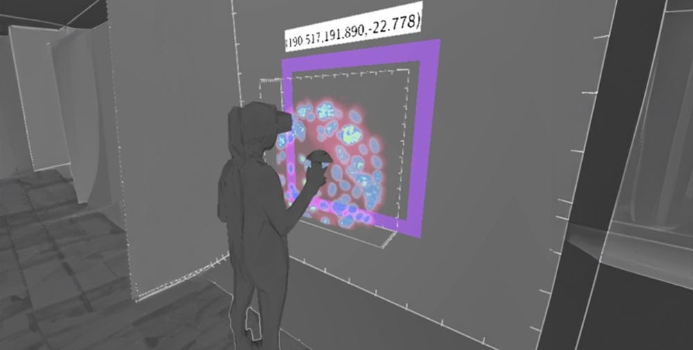
XR microscopy could lead to a wave of new insights here. For the first time, users can interact directly and in 3D with the sample under the microscope. This enables certain modifications to the living sample – such as laser ablation or bleaching experiments – to be carried out not only with better inclusion of the spatial environment of the cell, but also faster and more accurately than with a two-dimensional representation.
Virtual reality glasses instead of a monitor
The brains behind the concept are the two former CASUS scientists Dr. Ulrik Günther and Jan Tiemann. Prof. Ivo Sbalzarini from the Max Planck Institute of Molecular Cell Biology and Genetics (MPI-CBG) Dresden and Prof. Matthew McGinity from the Technical University of Dresden (TUD) are also part of the XR microscopy team. “The system we have designed does not require any specialized hardware,” says Günther. “It is initially based on virtual reality goggles and a standard computer running our software. We are developing interfaces for the major manufacturers of fluorescence microscopes as well as for important sample manipulation devices, so that XR microscopy will be possible on almost every existing modern research microscope in the near future.” The software is largely open source and will be developed further step by step together with the research community. Only for the interfaces to the microscopes and devices of commercial manufacturers will license fees be due.
“In our opinion, the XR microscopy approach offers clear added value,” says Prof. Otger Campàs from the TUD, who is one of the first to use an XR microscope set up by the developers. Campàs holds the Chair of Tissue Dynamics and is spokesperson for the Physics of Life Cluster of Excellence, which focuses on the investigation of three-dimensional biological data. “Thanks to XR microscopy, it is possible to work with complex data in a natural way. In principle, this technology enables meaningful experimentation with complex living samples in the first place”, Campàs continues.
On behalf of the HZDR Technology Transfer and Innovation Department, which supports the project team on the path from idea to product, Ascenion GmbH in Munich, an expert in knowledge and technology transfer in the biosciences and medicine, also took a close look at the project. Due to the high growth in the addressed markets and the interest of established players such as the microscopy companies Leica and Zeiss in the topic, Ascenion concluded that the project offers a promising opportunity for a successful spin-off.
After winning the innovation competition a year ago, Günther and Tiemann first submitted an application for the grant of a patent. In summer 2024, they then received validation funding from the Sächsische Aufbaubank. Thanks to the current BMBF funding via the GO-Bio initial program, the two specialists can explore the innovation potential of their idea, carry out market analyses and evaluate the patent situation. In the best-case scenario, the business idea will develop into a viable business model.
Contact
Dr. Ulrik Günther | Project Manager XR-Microscopy
Helmholtz-Zentrum Dresden-Rossendorf e. V. (HZDR)
Tel.: +49 3581 37523-52
E-mail: ulrik.guenther@hzdr.de
– – – – –
Further links
👉 www.hzdr.de
Photo: U. Günther & J. Tiemann/HZDR
